Posts

Event 3: Final Review/Study Guide – 5/24

Image
1. I attended the final review and signed out at the end of the session. 2.  Study Guide:         On May 24 th , I attended a review session for the DESMA (Design Media Arts) 9 course, an Art|Sci course that helped me create this blog revolving around the intersection of art, science, and technology. With only two weeks left in the Spring Quarter at UCLA, it was crucial for me to attend this two-hour, PowerPoint-based review at the Broad Arts building. It was led by Professor Victoria Vesna, as well as all of the teaching assistants who guided the students through this course. Following this review, I have created the following study guide to help me successfully complete this course. Most importantly, the study guide contains deadlines for the final project and blog summary assignments, as well as some guidelines and advice. Caption : The slide outlining important deadlines for DESMA 9, from the final review’s PowerPoint lecture.             The quiz and blog summary ass

Week 8: Nanotech + Art

Image
For this week’s blog post, I listened to James Gimzewski’s lectures on the history, advantages, and prevalence of nanotechnology. Previously a researcher at IBM’s Zurich Research Laboratory, Gimzewski is currently a chemistry professor and nanoscientist at UCLA. His online lectures gave me a new perspective on the reality of nanoparticles and nano-scale processes, which exist everywhere from nature to consumer products. Starting in the 1960s, people began approaching nanomaterials as potential for accomplishing unmet needs. Nanoscience grew in the 1980s, following the discovery of graphene and buckminsterfullerene (or the buckyball). Nanotechnology such as scanning tunneling microscopy (STM) and atomic force microscopy (AFM) provided scientists with the ability to visualize and even alter material surfaces on the atomic scale. Nanoparticles have much different properties compared with their bulk state, which opens countless possibilities for science, technology, and perhaps art. After

Week 7: Neuroscience + Art

Image
            This week, students of DESMA 9, a class in the Design | Media Arts department at UCLA taught by Victoria Vesna, learned about how neuroscience inspires artists who are fascinated with the human mind. The history of neuroscience is relatively short, starting with the theory of phrenology in 1796 by Franz Joseph Gall. This physician believed that various cerebral functions were localized, meaning that individual intellect and personality could be predicted based on brain or skull shape. Ramon y Cajal (1852-1934) then pioneered microscopic studies of the structure of the nervous system, receiving the Nobel Prize in 1906 for being the founder of neural anatomy. Most recently, the Brainbow was invented in 2007 by Harvard professors of neurobiology, which effectively tagged individual neurons in the brain with fluorescent proteins. This revolutionary technology led to beautiful and informative images for scientists (Vesna, 2012). (Complex neural pathways in a zebra fish la

Week 6: BioTech + Art

Image
            Just before the start of the 21 st century, a new type of art utilized genetic engineering to create unique, aesthetically intriguing organisms. Known as BioArt today, these art projects were pioneered by Joe Davis, and shortly after, Eduardo Kac. Joe Davis was both an artist and a researcher at MIT’s Center for Advanced Visual Studies. Although a rather eccentric human being, he set many precedents for future BioArtists. He realized that genes and genomes could become a new palette, with which artists can use life itself as a valid expressive medium. He theorized many projects, such as the modifying amino acids to form highly geometric capsules, but many molecular biologists were still hesitant to teach him all of the necessary techniques. Shortly after, Eduardo Kac invented the “GFP Bunny” in 2000, an iconic piece of BioArt (which he called transgenic art at the time). In short, he injected the Green Fluorescent Protein (GFP) into fertilized albino rabbit cells and grew

Event 2: Mnemoawari – by Elí Joteva

Image
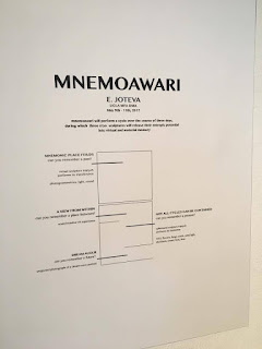
This past Tuesday, on May 9 th , I attended Elí Joteva’s solo exhibition, titled “Mnemoawari”, at the Broad Arts Center in UCLA. Currently a graduate student in UCLA’s Design | Media Arts department, Joteva is also an inter-media artist and researcher who combines neurophysics and art through the use of imaging tools and biofeedback technologies. In other words, she is interested in phenomena related to brain function and presents art pieces that incorporate bioimaging techniques. The first thing that intrigued me about this solo show was the title: mnemo- , meaning memory in Greek, and -awari , which translates to wanderer in Hindi. Walking into the gallery, I immediately saw a sign that described the purpose of her exhibition. It was a pleasure meeting Joteva at the exhibition, and I enjoyed experiencing the artist’s expression of memory and time! (Exhibition sign (left picture), and a hanging cryogenic sculpture and VR headset in the background. There were a lot of people

Kevin_Yin_DESMA9_Midterm

Link to PDF of my Midterm (Also submitted online through UC Canvas): https://drive.google.com/file/d/0B58o_vgxPHPtc3I3WjBDcUp3T2M/view?usp=sharing

Week 4: Medicine + Technology + Art

Image
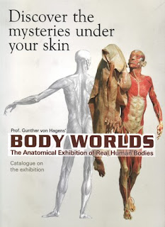
            The medical fields, particularly human anatomy, have always fascinated artists. Understanding human body structure components such as the skeleton and muscles is an essential skill for many art students. One modern example of how art and medicine intertwine is the BODY WORLDS exhibit, a touring art gallery that features perfectly preserved human organs and bodies. This special exhibition is in fact the product of a long history of medical technology. Specifically, Gunther von Hagens’s plastination technique (discovered in 1979) made it possible to preserve biological tissue specimen using polymer mixtures. This type of art reflects on society’s changing perceptions of human anatomy. From analyzing individual cadavers to creating databases on systems of microbial organisms in the body, medical technology continues to inspire revolutionary pieces of art (Vesna, 2012). (The book cover of Gunther Von Hagens's Body Worlds Catalog, first published in 2006.)